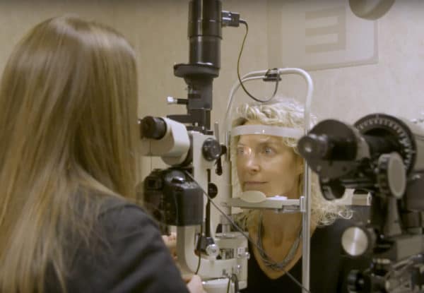
In addition to routine comprehensive eyecare, the doctors at Kirman Eye provide emergency eye care including removal of corneal foreign bodies, treatment of corneal abrasions, and conjunctivitis (“pink eye”). If you find yourself with an eye emergency or require 24 hour emergency eye care, we offer treatment for a host of chronic ocular diseases such as AMD, Dry Eye Syndrome, and Glaucoma is also available at Kirman Eye. Some of our special tests include:
- Dark Adaptation – The AdaptDx instrument allows your doctor to diagnose Age-Related Macular Degeneration (AMD) up to three years before any changes in the macula can be seen or any vision loss occurs. Kirman Eye doctors have been using this technology since 2014 to help diagnose AMD and then design a treatment plan to help patients maintain their vision.
“The AdaptDx is the most significant diagnostic breakthrough in my lifetime to help doctors manage the leading cause of blindness in the United States.” – Dr. Kirman
- OCT – Optical Coherence Tomography allows your doctor to view an “optical cross-section” of the retina and choroid layers of the back of your eye. This technology is very useful in detecting small abnormalities in the retinal layers that can interfere with good vision such as AMD or macular edema from Diabetes. The OCT has been the successor of HRT technology at analyzing the contour of the optic nerve and ganglion cell layer health which can be affected by glaucoma and other optic nerve disorders.
- Visual Fields – Visual field testing allows your doctor to understand your ability to see centrally as well as your peripheral vision in each eye. Diseases and events such as Glaucoma, Multiple Sclerosis, Strokes, Concussion, & Lyme may affect your visual field in one or both eyes.
- Optomap – The Optomap retinal imaging device allows your doctor to view a panoramic image of the retina to look for abnormalities caused by Diabetes, Stroke, Glaucoma, Retinal Detachments, Holes, Tears, Cholesterol, and Hypertension. Because each eye is unique, the image does a much better job of documenting the retinal condition compared to notes or diagrams drawn by the doctor. Optomap imaging documents a baseline of the retina which is then used in future years to compare and look for subtle changes that would alert your doctor to a potentially sight-threatening condition. It is also true telemedicine in that the images can be transferred to another doctor anywhere in the world using the same technology.
- Quantify (MPOD) – The Quantify instrument measures the Macular Pigment Optical Density (MPOD) of each retina. The macula is responsible for our central vision and is filled with pigments (Lutein & Zeaxanthin) that are essential for proper function and protection of the rod and cone cells. If the macular pigment is found below, there is not as much protection from Ultra Violet light for the rod and cone cells. Low Macular Pigment Optical Density is a risk factor for macular diseases like AMD. MPOD levels can be improved by taking quality vitamin supplements that are high in Lutein & Zeaxanthin
- Pachymetry – This is the measurement of the thickness of your central cornea (the front surface of the eye). This measurement was found to be critically useful for managing patients with a suspicion of glaucoma. Thinner corneas have a tendency to give lower intraocular pressure readings while thicker corneas give higher intraocular pressure readings (Glaucoma test). This measurement is used along with other diagnostic tests for the diagnosis of Glaucoma in a timely matter. It is also used to monitor corneal conditions such as Keratoconus and other corneal dystrophies that can cause visual blur.


