ADAPTDX
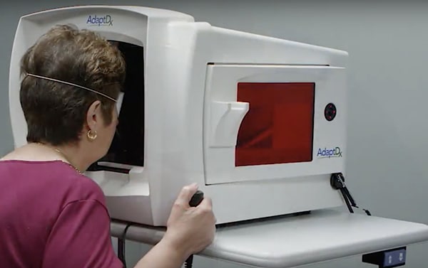
The AdaptDx instrument is used to objectively measure retinal function based on dark adaptation (recovery of visual function when going from light to darkness). Patients with early stages of age related macular degeneration (AMD), retinitis pigmentosa (RP), and other diseases of the retina can have reduced dark adaptation function. This is an “early warning device” in predicting your risk of future AMD. This valuable data will help your doctor assess your risk for AMD, RP, and other retinal diseases. The AdaptDx is helpful in early detection of problems that may not be found with other instruments until later in the disease process when vision has already been affected.
Testing Time Frame: Varies by individual, may take up to an hour
OPTICAL COHERENCE TOMOGRAPHY
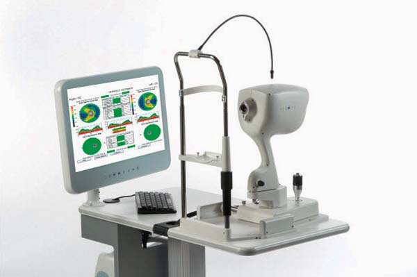
Optical coherence tomography (OCT) is a non-invasive imaging test. OCT uses light waves to take cross-section pictures of your retina.
With OCT, we can see each of the retina’s distinctive layers. This allows our optometrists to map them and measure their thickness. These measurements aid in the diagnosis and monitoring of various diseases such as age-related macular degeneration, and diabetic eye disease. They also provide treatment guidance for glaucoma and diseases of the retina.
Testing Time Frame: Varies by the individual, scanning usually takes about 5 – 15 minutes.
QUANTIFEYE
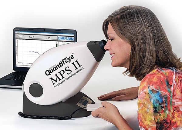
The QuantifEye instrument measures Macular Pigment Optical Density (MPOD) in the retina (back of the eye). If you think of the retina as a target, the macular area is the “bullseye.” The macula is responsible for our central vision (20/20 vision) and most of our color vision. Normal macular pigment levels effectively block harmful blue light from reaching the visual cells in the macular area, reducing the risk of damage to your central vision. Low macular pigment levels are highly associated with age-related macular degeneration (AMD) and poor night vision performance such as glare and haloing while driving at night.
This device determines your MPOD score which helps your doctor to assess your risk of age-related macular degeneration as well as your degree of decreased night vision performance. This MPOD measurement also helps to determine whether you are a candidate for dietary supplements for the eye. Specialized eye supplements are recommended by your doctor to increase your MPOD score to lower your risk of developing AMD. The supplements can also improve night vision performance.
Testing Time Frame: 10-15 minutes
OPTOMAP
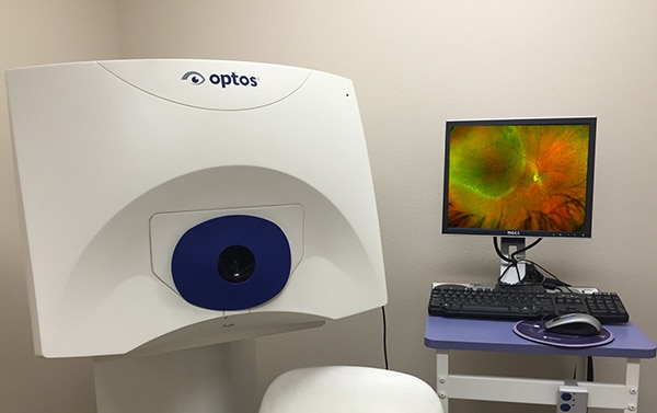
The Optomap® retinal image gives the Kirman Eye professionals a much larger view (200 degrees) of the back of the eye – your retina – than conventional eye exam equipment. The images can be taken without dilating your pupils which is a very common procedure that is uncomfortable and inconvenient for many people. Each Optomap® image is as individual as fingerprints or DNA and can provide eye care professionals with a unique view of your health very quickly and comfortably. The Optomap® image is captured in less than one second and is immediately available for you and your doctor to review.
Testing Time Frame: Less than 5 minutes
VISUAL FIELD
Diseases like glaucoma, age-related macular degeneration, multiple sclerosis, stroke, and brain tumors can cause defects in your visual field. Visual field testing is used to monitor the effectiveness of glaucoma treatment, the progression of optic atrophy due to multiple sclerosis, improving or worsening visual effects of a stroke, and other visual pathway disorders over time.
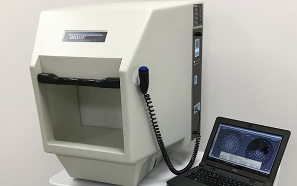
The Dicon Visual Field Instrument tests your ability to see varying intensities of light throughout your central and peripheral (side) vision in each eye. This allows our doctors to assess the health of your visual pathway from the retina (back of the eye) through each side of the brain to the visual center in the back of your brain. The equipment uses advanced software to map the area of vision in each eye. The results of the test allow your doctor to determine the sensitivity of your central and side vision.
Testing Time Frame: about 30 minutes
Our newest addition to the office, the LipiScan Imager and LipiFlow treatment system are helping our practice to address dry eye issues in a whole new way!
LIPISCAN DYNAMIC MEIBOMIAN IMAGER (DMI)
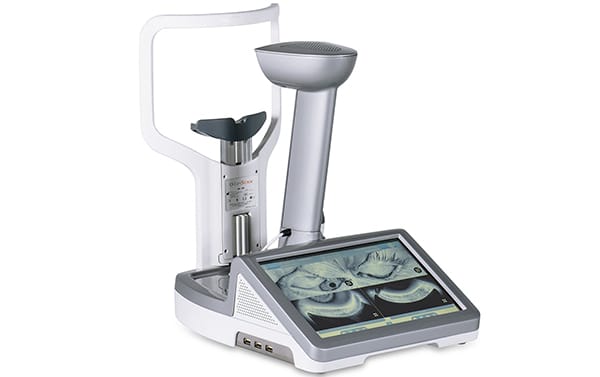
The LipiScan Imager allows Kirman Eye professionals to check for signs of Meibomian Gland Dysfunction (MGD) which can cause dry eye syndrome. Meibomian glands secrete an oily substance which coats the tear film covering the eye and prevents evaporation of your tears. A healthy tear film keeps our eyes nourished and comfortable. LipiScan gives our doctors a high definition image that shows the health of your meibomian glands. This allows them to choose the most appropriate treatment for each patient’s unique needs.
Testing Time Frame: 2 minutes
LIPIFLOW
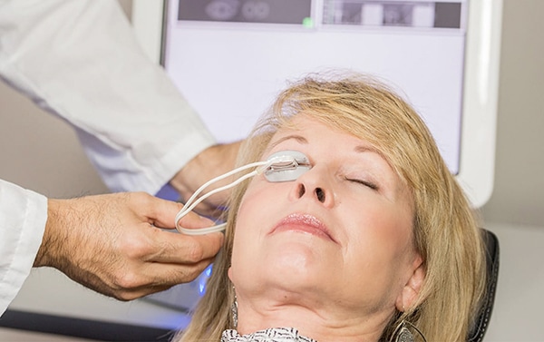
LipiFlow is an FDA approved treatment for Dry Eye Syndrome caused by meibomian gland dysfunction. LipiFlow gently massages the outer lid while treating the glands on the inner lid by applying low heat. This helps to remove any blockages from the meibomian glands which allow them to produce the oils that make up the top protective oil layer of the tear film.
This is a “dropless” treatment for dry eye and, in many of our patients, reduces the inconvenience of daily eye drops. Results vary by patient, however, most patients see a significant increase in their comfort and quality of life. LipiFlow treatments are done in-office and only take 12 minutes to complete. However the patient should expect to be in the office 30-45 minutes for pre-treatment and post-treatment observation. Both eyes are treated at the same time in order to reduce the time a patient has to spend at our office.
Testing Time Frame: Varies by patient

